SERVICES
Ultrasonography Radiology MRI CT Education
Mobile ultrasonography service to practices in the South West of England
|
Radiographic reporting of digital studies submitted directly over the internet
|
MRI studies reported using simple online file transfer service
|
CT study reporting via simple online file transfer service
|
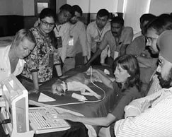
Formal lecturing, seminars, and practical tuition in ultrasonography and radiology
|
ULTRASONOGRAPHY

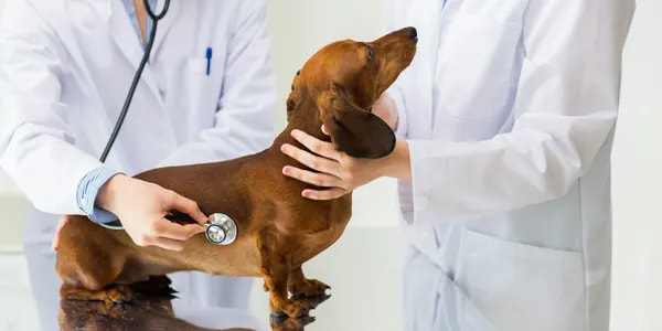
Ultrasonography Service
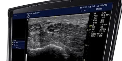
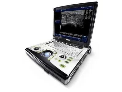
We offer a mobile ultrasonography service to practices in the South West of England using advanced portable ultrasound equipment.
We offer a mobile ultrasonography service to practices in the South West of England using advanced portable ultrasound equipment.
|
What is Ultrasound?
Ultrasonography is an invaluable and under-used tool in the diagnosis of many disease conditions. The use of ultrasound imaging provides important diagnostic information as to the nature of thoracic, abdominal and musculo-skeletal pathology, which may not be evident from other imaging modalities such as radiography. |
|

|
Indications
|
|
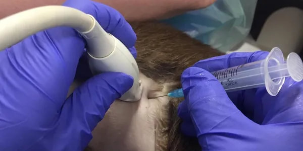
Sampling
Ultrasonography can be used to perform ultrasound-guided procedures, such as tissue core biopsy, fine needle aspiration and cystocentesis. The use of these techniques can reduce the degree of stress and discomfort experienced by the patient, compared to a more invasive technique such as laparotomy. |
|
RADIOLOGY
|
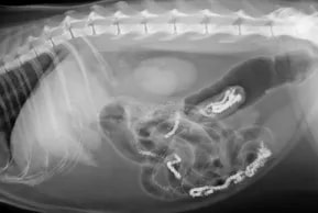
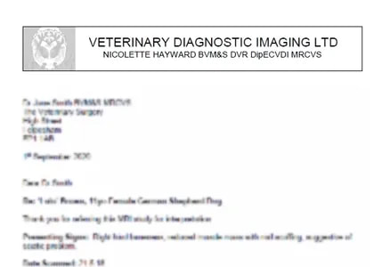
Reporting
Radiographic reports include the following information:
|
|
Submissions
DICOM files can be transferred via our Image Upload System |

|
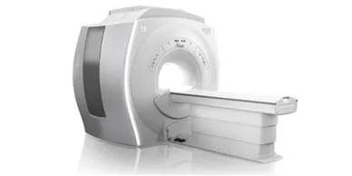
MRI
What is MRI? Indications Submissions
|

|

|
|
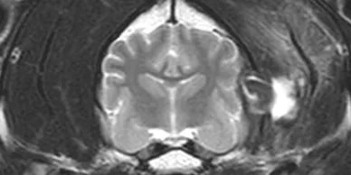
Whilst imaging of the brain and spinal cord have been the most common indications for MRI in recent years, the list of conditions where MRI has proven invaluable grows rapidly.
MRI has several advantages over other forms of imaging. This modality does not require the use of ionising radiation, and is non-invasive, unlike myelography. MRI can obtain images in any plane, and provides excellent contrast resolution, offering soft tissue information in exquisite detail. Oedema and inflammation, which are commonly associated with pathological change can be detected in soft tissues, but also within bone. MRI is a highly useful tool in surgical planning, enabling the surgeon to ascertain the extent of pathology e.g. tumour removal, and thus the margins required for adequate resection. |
|
DICOM files can be transferred using our Image Upload System
|
CT

|
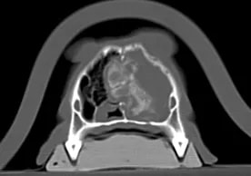
What is CT?
The availability of Computed Tomography is becoming more widespread, and this form of cross-sectional imaging has numerous veterinary applications. There is considerable crossover between the indications for CT and MRI, although the identification of certain types of pathology is more typically associated with CT. For example, high resolution CT has a much higher sensitivity for small pulmonary metastatic lesions than either conventional radiography or MRI. Like MRI, CT is invaluable in determining the scale of pathology, characterising lesions to optimise surgical planning. |
|
Indications
|
|

Submissions
DICOM files can be transferred via our Image Upload System
DICOM files can be transferred via our Image Upload System